")
SELECTIVE BLOCKADE OF CaV1.2 VERSUS CaV1.3 L-TYPE Ca2+ CHANNELS BY THE BLACK MAMBA TOXIN CALCISEPTINE
INHIBITION SPÉCIFIQUE DES CANAUX CALCIQUES DE TYPE L CaV1.2 VERSUS CaV1.3 PAR LA TOXINE CALCISEPTINE ISSUE DU VENIN DE MAMBA NOIR
Les canaux calciques de type L sont impliqués dans de nombreuses fonctions physiologiques. Les cellules myocardiques expriment deux isoformes de canaux L à savoir Cav1.2 et Cav1.3. Cav1.2 est exprimé de façon ubiquitaire dans les tissus cardiaques ventriculaires et supraventriculaires incluant les nœuds sinoatrial (SAN) et atrioventriculaires (AVN). Cav1.2 est l’isoforme prédominante dans le ventricule adulte où il est responsable du couplage excitation-contraction et constitue la cible de la voie AMP- dépendante qui médie l’effet inotrope positif des catécholamines sur la contraction cardiaque. Cav1.3 est exprimé dans les oreillettes et en particulier fortement exprimé dans les cellules du SAN, du AVN et du tissu conducteur. Ainsi Cav1.3 joue un rôle majeur dans l’activité pacemaker du SAN et la conduction de l’influx dans le AVN. Malgré l’importance d’identifier les rôles physiologiques respectifs de Cav1.2 et Cav1.3 dans les tissus cardiaques mais aussi dans les cellules neuronales, il n’existe pas à l’heure actuelle d’outil pharmacologique sélectif disponible. Les venins animaux constituent un immense réservoir pour l’identification des toxines capables d’inhiber spécifiquement des isoformes de canaux ioniques et déterminer leur fonction dans divers tissus.
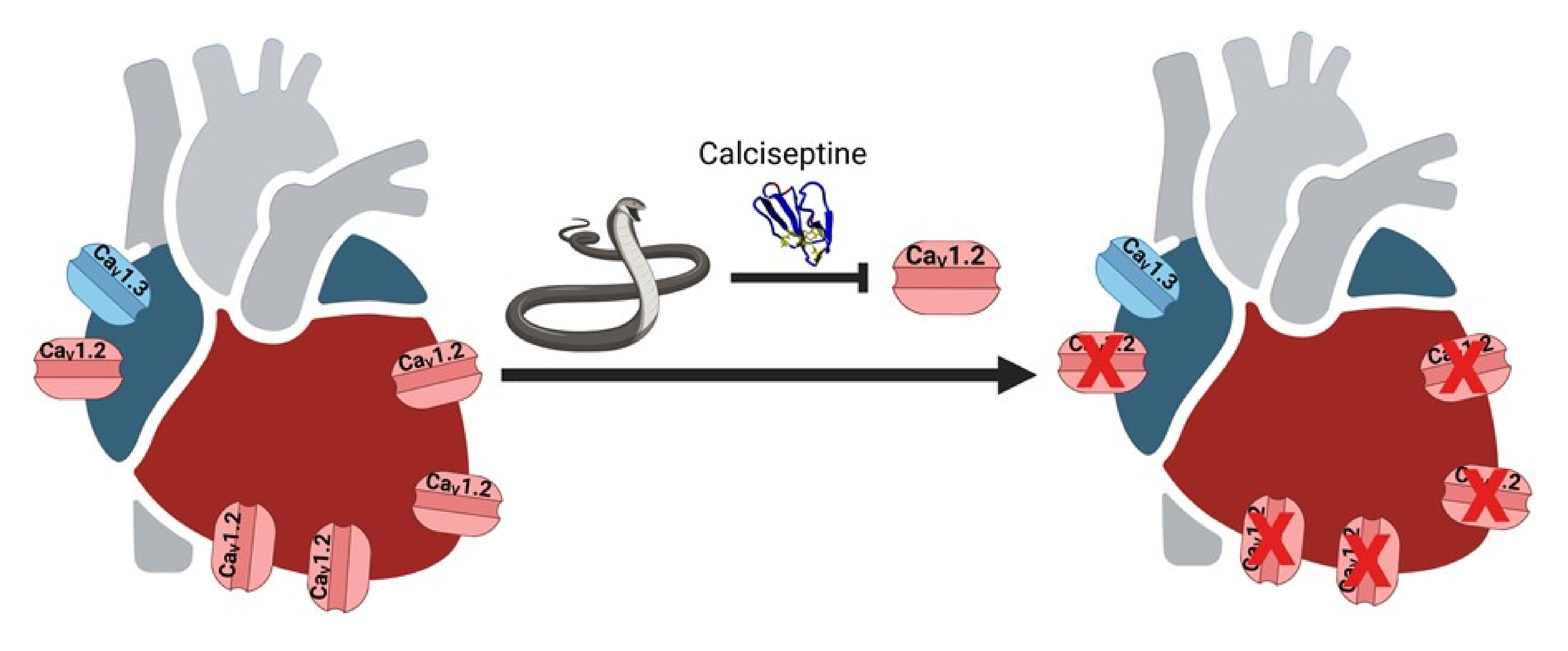
Un travail collaboratif impliquant trois équipes de l'IGF (équipe "Cardioprotection, physiopathologie du rythme cardiaque et ischémie" animée par Stéphanie Barrère et Matteo Mangoni, équipe "Dynamique cellulaire des canaux calciques et nociception" animée par Emmanuel Bourinet, équipe "Canaux Ioniques-Excitabilité neuronale et canalopathies" animée par Philippe Lory), l'Institut de Pharmacologie Moléculaire et Cellulaire à Valbonne (Université Côte d'Azur) ainsi que le Département de Physiologie de l'Université Nationale de Singapour, a permis de montrer que la calciseptine (Cas), un polypeptide issu du venin du mamba noir, bloque sélectivement les canaux Cav1.2, sans affecter les canaux Cav1.3. Cas diminue puissamment la contractilité cardiaque sans affecter la fréquence cardiaque d'un cœur de souris perfusé à Langendorff. Alors que Cas bloque totalement ICaL (Cav1.2) dans les myocytes ventriculaires, il inhibe partiellement le courant ICaL (ICav1.2+ICav1.3) dans les myocytes SAN de souris témoins mais bloque totalement ICaL dans les myocytes des souris Cav1.3-/-, où seul Cav1.2 est fonctionnel. La sélectivité du blocage par Cas de Cav1.2 par rapport à d'autres canaux calciques a été confirmée dans des HEK-293T exprimant des canaux recombinants Cav1.2, Cav1.3 (variants d'épissage Cav1.3 42a et Cav1.3 42), Cav3.1 (type T,) Cav2.2 (type N) et Cav2.1 (type P/Q).
En conclusion, cette étude montre que la calciseptine bloque sélectivement les canaux Cav1.2 dans le cœur et représente un outil important dans le panel des modulateurs des canaux calciques pour aider à disséquer le rôle de ces deux isoformes des canaux calciques L dans la physiopathologie cardiaque, mais aussi dans les cellules neuronales où chaque isoforme est décrite comme médiant des fonctions physiologiques spécifiques.
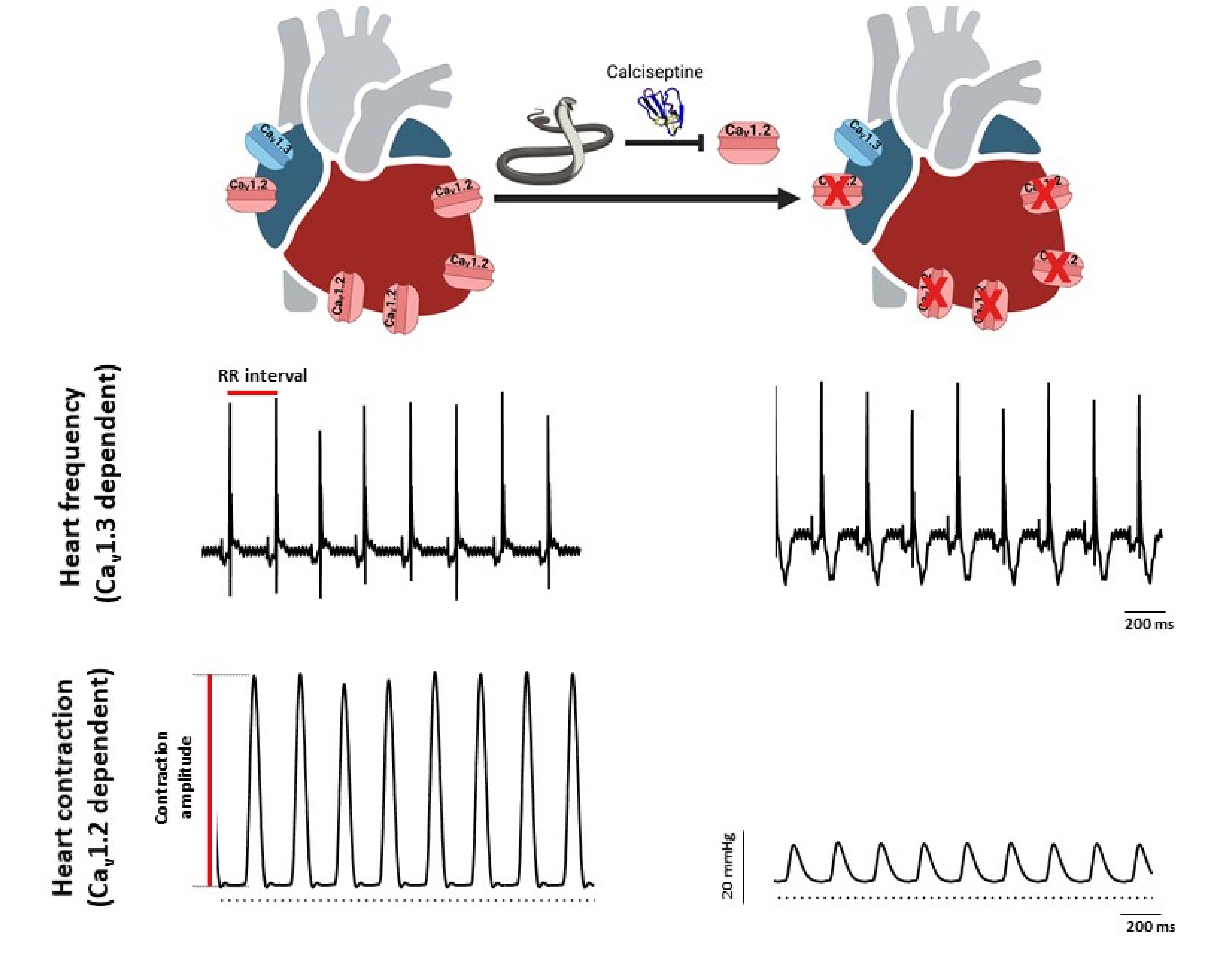
Fréquence cardiaque et contraction ventriculaire dans le cœur perfusé en présence de calciseptine. Tracés ECG (haut) et enregistrements de pression (bas) d'un cœur isolé perfusé, en condition contrôle (gauche) et en présence de calciseptine (droite).
...................................................................................................................................................................................................................................................................................................................................................................
SELECTIVE BLOCKADE OF CaV1.2 VERSUS CaV1.3 L-TYPE Ca2+ CHANNELS BY THE BLACK MAMBA TOXIN CALCISEPTINE
L-type voltage-gated calcium channels are involved in multiple physiological functions. The heart expresses two L-type Ca2+ channels isoforms, Cav1.2 and Cav1.3. Cav1.2 is ubiquitously expressed in the heart ventricular and supraventricular chambers, as well as in the sinoatrial (SAN) and atrioventricular (AVN) nodes. Cav1.2 is the predominant L-type isoform in adult ventricle, where it couples excitation to contraction and constitutes the target of the cAMP/PKA mediated positive inotropic effect on cardiac contractility exerted by catecholamines. Cav1.3 is expressed in the atria and is strongly expressed in the SAN and AVN. Cav1.3 plays a major role in SAN pacemaker activity and impulse conduction through the AVN. Despite the importance of dissecting the physiopathological roles of Cav1.2 and Cav1.3 channels in cardiac myocytes and neuronal cells, no selective pharmacological tool is currently available. Animal venoms constitute an immense reservoir for identification of new toxins and toxin-derived tools for identifying the physiological function of ion channels in various tissues.
A collaborative study involving three IGF teams (team "Cardioprotection, pathophysiology of heart rhythm and ischemia" led by Stéphanie Barrère and Matteo Mangoni, team "Calcium channel dynamics and nociception" led by Emmanuel Bourinet and team "Ion channels in neuronal excitability and diseases" led by Philippe Lory), the Institute of Molecular and Cellular Pharmacology (Valbonne, Université Côte d'Azur) and the Department of Physiology at the National University of Singapore, has shown that Calciseptine (Cas), a polypeptide from the venom of the black mamba, selectively blocks Cav1.2 channels, without affecting Cav1.3 channels. Cas potently decreases cardiac contractility without affecting heart rate of Langendorff perfused mouse heart. While Cas totally blocks ICaL (Cav1.2) in ventricular myocytes, it partially inhibits ICaL current (Cav1.2+Cav1.3) in SAN myocytes from wildtype mice and fully blocks ICaL in myocytes from Cav1.3−/− counterparts, where only Cav1.2 is functional. The selectivity of Cas blockade of Cav1.2 versus other calcium channels has been confirmed in HEK-293T expressing recombinant Cav1.2, Cav1.3 (both Cav1.342a and Cav1.342 splice variants), T-type Cav3.1, N-type Cav2.2 and P/Q-type Cav2.1 channels.
In conclusion, this study shows that Cas selectively blocks Cav1.2 channels in the heart, providing an important tool in the panel of calcium channel modulators to help dissect the role of this channel isoform in heart physiology and automaticity but also in neuronal cells in which each isoform is mediating specific physiological functions.
Heart rate and ventricular contraction in Cas perfused heart. Representative ECG traces (top) and pressure recordings (bottom) from isolated perfused heart in control condition (left) and in the presence of calciseptine (right).