")
UN RÉSEAU NEURAL MÉDIAL POUR LE DÉPLOIEMENT VOLONTAIRE DE L’ATTENTION VISUO-SPATIALE
L’évaluation des patients souffrant d’une négligence spatiale unilatérale gauche, un syndrome neuropsychologique communément observé après une lésion neurologique de l’hémisphère droit, et qui se caractérise principalement par un défaut d’exploration du champ visuel gauche ou une perte de conscience des évènements qui s’y produisent, a permis des avancées majeures dans notre compréhension des systèmes neuronaux impliqués dans l’orientation de l’attention visuo-spatiale. Toutefois, l’implication des structures situées sur la face médiale du cerveau dans cette fonction centrale pour l’organisation des comportements guidés visuellement n’avait pas encore été clairement établie car l’ensemble des études disponibles se reposait très largement sur l’accident vasculaire cérébral comme modèle lésionnel – une condition neurologique que ne touche que très occasionnellement ces structures.
Dans cette étude réalisée par deux membres de l’Institut de Génomique Fonctionnelle (CNRS/Inserm/Université de Montpellier), Guillaume Herbet et Hugues Duffau, et publiée récemment dans le journal Nature Communications, il est démontré que deux aires cérébrales logées dans la partie médiale du cortex frontal, les champs oculaires supplémentaire et cingulaire, ainsi que leur connectivité de substance blanche sous-jacente, contribuent au déploiement de l’attention visuo-spatiale, puisqu’une lésion de ces structures cause une forme très spécifique de négligence spatiale (la négligence d’exploration visuo-motrice) caractérisée par un déficit de l’exploration volontaire du champ visuel gauche sans perte de conscience de celui-ci.
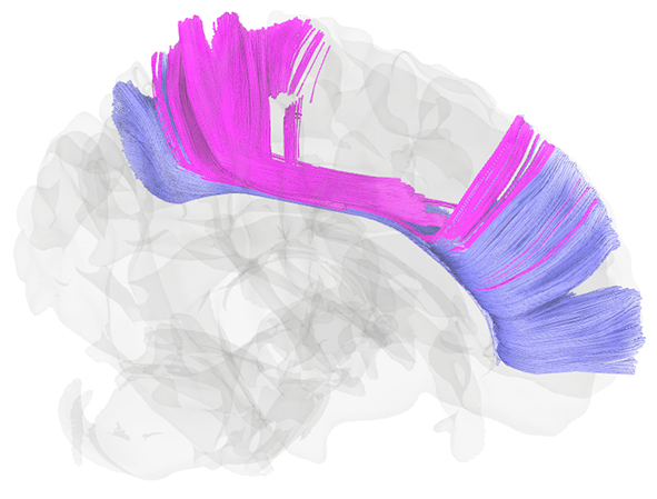
Pour aboutir à ce résultat, les deux chercheurs se sont appuyés sur l’évaluation comportementale et longitudinale (c.-à-d. avant et après la chirurgie) de nombreux patients souffrant d’un gliome de bas grade, une tumeur rare du système nerveux central qui affecte fréquemment les régions mésiales antérieures. Des corrélations anatomo-fonctionnelles ont été réalisées en combinant des méthodes de cartographie lésion-déficit basée sur l’apprentissage automatique et d’analyse des déconnexions structurelles. Les résultats ont montré que l’exérèse chirurgicale des champs occulaires médiaux perturbait très significativement l’exploration spatiale du côté gauche au cours d’une tâche de recherche visuelle dans l’après-chirurgie, au même titre que l’atteinte de la branche I du faisceau longitudinal supérieur, une connexion de substance blanche associative interconnectant les cortex frontal et pariétal médiaux. Ces résultats devraient avoir une implication importante pour les modèles neurocognitifs et neuro-computationnels de l’attention visuo-spatiale.
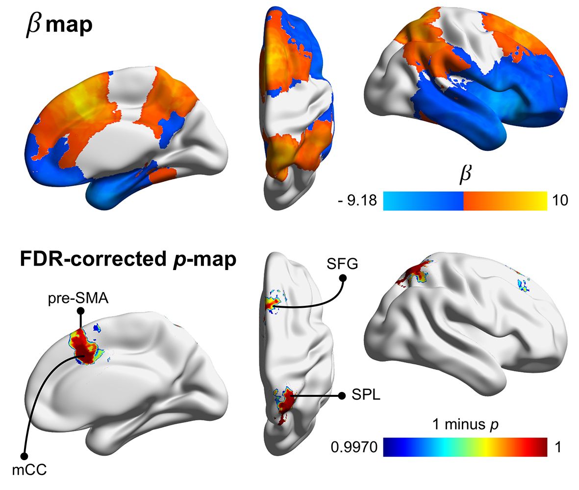
Cartographie spatiale de la relation statistique entre la localisation des résections chirurgicales et la négligence d’exploration visuo-motrice.
Spatial mapping of the statistical relationship between the location of surgical resections and visuo-motor exploratory neglect.
.....................................................................................................................................................................................................................................................................................................................................................................................
A MEDIAL NEURAL NETWORK FOR THE VOLUNTARY DEPLOYMENT VISUO-SPATIAL ATTENTION
The assessment of patients suffering from unilateral spatial neglect, a common neuropsychological syndrome caused by a neurological damage of the right cerebral hemisphere and characterized by difficulties in exploring the left visual field or by a loss of awareness of stimuli occurring in this field, has allowed important advances in our understanding of the neural networks involved in the orientation of visuo-spatial attention. However, the precise contribution of structures shaping the medial face of the brain to this function vital for visually-guided behaviors was to date not clearly established, mainly because available studies were mostly based on vascular injuries as lesion model – a common neurological condition that affects the medial structures very occasionally.
In this study conducted by two researchers from the Institute of Functional Genomics (CNRS/INSERM/Université de Montpellier), Guillaume Herbet and Hugues Duffau, and recently published in the journal Nature Communications, it is shown that two cerebral areas lodged in the medial part of the frontal cortex, the supplementary and the cingular eye fields, as well as their underlying white matter connectivity, contribute to the voluntary deployment of visuo-spatial attention, as damage to these structures causes a specific form of spatial neglect (visuo-motor exploratory neglect) characterized by a deficit in exploring the left visual field without loss of consciousness of it.
To achieve this finding, the researchers relied on the longitudinal (i.e. before and after surgery) behavioral assessment of a large sample of patients harboring a low-grade glioma, a rare tumor of the central nervous system that frequently affects the medial frontal cortex. Anatomo-functional correlations were performed by combining machine learning-based lesion-deficit mapping and structural disconnection analyses. The obtained results showed that surgical removal of the medial eye fields disturbed the voluntary exploration of the left visual scene during a visual search paradigm after surgery, as well as damage to the branch I of the superior longitudinal fasciculus – an associative white matter tract interconnecting the medial part of the frontal and parietal cortex. These results have important implications for current neurocognitive and neuro-computational models of visuo-spatial attention.